WINSTON-SALEM, NC – A young Gastroenterologist and his team made history last week when what was supposed to be a standard endoscopic exam turned into a brave push towards the unexplored frontiers of medical science. Gastroenterologists of Salem General Hospital described a revolutionary new technique that they termed “Esophagogastroduodenojejunoileocolonoscopy” in a press release this afternoon. Widely regarded as a ‘maximally-invasive procedure’, this operation requires a regular endoscope, about 30 feet of tube, and an eager practitioner with a complete lack of common sense.
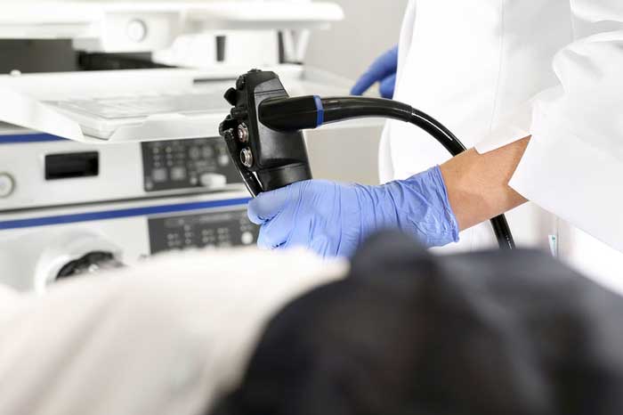
It all began on a Thursday morning when Gastroenterology fellow Dr. Richard Baum-Fondler began performing a routine endoscopic exam on a middle-aged male patient. “I was only trying to find the source of a GI bleed,” said the humble young doctor. “The digital rectal exam and colonoscopy were inconclusive, so we decided to run a quick Esophagogastroduodenoscopy to see if we could spot the lesion.” he explained as he cracked his knuckles. “He just kept going and going,” recalled Endoscopy Suite nurse Alexandra Kelley, RN. “We were all watching the clock and waiting for him to finish up, but Dr. Richard went deeper than we ever thought possible.”
Dr. Baum-Fondler seemed lost in thought for a moment, as if trying to remember all the details of that fateful day. “The esophagus and stomach were clean, as we expected. Then I entered the duodenum to take a quick look, at which point I saw something that seemed like a polyp further ahead. I decided to try and maneuver the endoscope past that tight corner.” he explained.
“Doctor Richard was spinning that joystick and mashing all the buttons like crazy,” stated Jake Benally, a 3rd year medical student who was there to witness the historic moment. “The nurses seemed horrified but I think it turned out okay.” he concluded.
According to the operative note, it took the rookie gastroenterologist a total of 54 minutes to painstakingly navigate the patient’s entire GI tract. Several endoscopy tubes had to be connected end to end as he ventured boldly into the farthest reaches of the small intestine. “I didn’t find any polyps in the duodenum or the jejunum, but I wasn’t going to let that stop me.” remarked Dr. Baum-Fondler. “I kept running out of tube but we managed to connect new ones as we went, and eventually we encountered the ileocecal valve. I decided to keep going and take another look at the colon. You see, I wasn’t going to give up after going all that distance.” he said. The procedure was concluded after ‘a white structure with the appearance of fabric’ was encountered, which turned out to be the fully-clothed patient’s undergarments. At that point the patient was shifted to the twerk position and fully exposed to retrieve the endoscope. Operative note states that the upper end of the endoscopy tube was disconnected in order to accomplish this, and the medical student was instructed to grab the lower end and “yank it like you’re starting a lawnmower”.
Dr. Omar Al-Aswad, division head of Gastroenterology at Salem General Hospital noted that there were no intraoperative complications and added, “This groundbreaking approach has wide-ranging implications for the future of rectal health. We are already planning to employ this technique in support of the new ACG guidelines that recommend flossing your rectum daily.”


