
Contrary to popular opinion, the eye exam can be performed by other health care providers too, not just ophthalmologists. Also contrary to popular opinion, “ophthalmology” is spelled with two H’s, not one or three. Before we dive into the eye exam, here are two reminders: (1) have the patient change into a gown before performing an eye exam since it may get messy, and (2) find an ophthalmoscope. An ophthalmoscope is an instrument used to inspect several parts of the eye and can be found in most pockets of eager medical students.
External Exam
Orient yourself; it can be embarrassing to perform a complete eye exam on a patient’s buttocks. Count the hairs in each eyebrow. Smell the eyelids. Taste the conjunctiva. Next, count the eyes: does the patient have one eye suggesting a cyclops or minion? Step back again: is the patient wearing glasses? goggles? an Iron Man helmet? Have the patient remove his or her Spiderman mask. Gaze deeply into your patient’s eyes and determine if it really is love at first sight. Feel free to sigh or smile shyly. Finally, spot the eye of the tiger.
Peekaboo
Visual Acuity
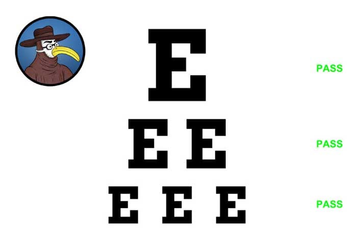
Have the patient stand 20 feet from you. With one eye covered, have the patient read the end credits of ABC’s 20/20. If the patient succeeds, he or she has 20/20 vision, which by the law of fractions equals 1. If you don’t have a TV handy, substitute in a Snellen eye chart. There are two Snellen eye charts: (1) the original Snellen, which has different letters of different sizes with a given visual acuity assigned to each line, and (2) the simplified Snellen (see picture above), which has only the letter E in different sizes with a given visual acuity of “Pass” assigned to each line.
Visual Fields
Have the patient look into your eyes. With your hands fully extended, flash your middle fingers, moving the birds around 360 degrees. If the patient is offended throughout the exam, then visual fields are normal.
Pupils
Observe the pupils: size? shape? symmetry? sexy? Anisocoria means something, as does miosis and mydriasis. Say the words “Argyll Robertson” out loud and see if the pupils react. Turn off the lights, bring out the candles, put on some Miles Davis, and shine your favorite penlight app into each pupil since regular penlights really, really suck. Note the direct reaction, consensual reaction, sensual reaction, and sexual reaction.
Extraocular Muscles
Have the patient follow your finger, pen, or sandwich with his or her eyes in the cardinal directions of gaze: up, up, down, down, left, right, left, right, B, A, Start. You have performed this exam correctly if you now have 30 lives.
Bedside Ophthalmoscopic Exam
Using the ophthalmoscope may seem awkward; it totally is. But it is important to perform a retinal exam now and again to destroy your self-esteem and reinforce your gratitude towards ophthalmologists. With the ophthalmoscope in your right hand, examine the patient’s right eye by leaning in and pretending to kiss him or her. A successful eye exam might involve the exchange of saliva. Note the red reflex: when ineptitude during this exam leads to the examiner blushing. Fail to identify the optic desk, retina, fovea, macula, and papilledema. However, if someone asks you if you can see them, enthusiastically reply, “Of course!”
Dilated Ophthalmoscopic Exam
An ophthalmologist will use special eye drops to help dilate the pupil. When the pupils are fully dilated at 10 cm, labor begins. Breech eye exams are tricky. Next, the ophthalmologist examines the retina for any signs of disease or placenta. Finally, the ophthalmologist patiently counts the number of rods and cones in each eye. Twice.
Intraocular Pressure
Tonometry tests for glaucoma by measuring IOP (intraocular pressure), which is not to be mistaken for OPP (yeah you know me). One can test for IOP grossly by pressing on the eyeball with the eyelid closed. If the eye bursts, you pushed too hard.
Refraction Testing
Refraction testing not only helps identify presbyopia, myopia, cornucopia, and onomatopoeia, but also helps spot the colors of the rainbow. When refraction testing is successful, an ophthalmologist can spot a pot of gold.
Slit Lamp Exam
The slit lamp is a microscope with a high-intensity light source to help an ophthalmologist (1) evaluate the eye with 3D views and (2) control your mind. Do not be surprised if you cluck like a chicken during this part of the exam.
Genital Exam
Here’s the part where it might get messy. Perform a thorough genital exam. You never know when those oculogenital fistulas will strike.
More Physical Exam Tips below:
– The Heart
– The Lungs
– The Abdomen
– The Nervous System


